

Scatter radiation is an inevitable by - product in medical imaging, which can significantly degrade image quality and increase the radiation dose to patients and medical staff. Tungsten, with its unique physical properties, plays a crucial and multi - faceted role in reducing scatter radiation in medical imaging. As a supplier of Tungsten for Medical Imaging, I have witnessed firsthand the importance of this remarkable metal in the medical field.
Physical Properties of Tungsten that Enable Scatter Reduction
Tungsten is a heavy metal with a high atomic number (Z = 74). This high atomic number is of great significance in the interaction with X - rays, which are commonly used in medical imaging such as radiography, fluoroscopy, and computed tomography (CT). When X - rays interact with matter, they can undergo different processes, including photoelectric absorption, Compton scattering, and pair production.
In the context of scatter reduction, the photoelectric effect and Compton scattering are the most relevant. The probability of the photoelectric effect is proportional to (Z^3/E^3), where (Z) is the atomic number of the absorber and (E) is the energy of the incident X - ray photon. Due to its high atomic number, tungsten has a relatively high probability of photoelectric absorption for low - to - medium energy X - rays. This means that when X - rays pass through a tungsten - based component, a significant number of photons are absorbed rather than scattered.
Compton scattering occurs when an incident X - ray photon interacts with an outer - shell electron of an atom, resulting in a change in the photon's direction and a reduction in its energy. Tungsten's high density ((\rho= 19.25\ g/cm^3)) also contributes to its effectiveness in reducing scatter. A higher density means that there are more atoms per unit volume for the X - rays to interact with. As a result, more Compton scattering events are likely to occur within the tungsten material, and the scattered photons are more likely to be absorbed by subsequent interactions with other tungsten atoms before they can reach the image receptor.
Applications of Tungsten in Scatter Reduction Devices
Anti - Scatter Grids
Anti - scatter grids are one of the most common devices used in radiography to reduce scatter radiation. They consist of alternating strips of an X - ray absorbing material and a radiolucent material. Tungsten is an ideal choice for the absorbing strips. The design of the grid is such that it allows primary X - rays (those that pass directly through the patient without scattering) to reach the image receptor while absorbing a large portion of the scattered X - rays.
The high absorption efficiency of tungsten in anti - scatter grids helps to improve the contrast of the radiographic image. By reducing the amount of scatter reaching the detector, the image becomes sharper, and the details of the anatomical structures are more clearly visible. This is particularly important in applications such as chest radiography, where accurate visualization of the lungs and other thoracic structures is essential for diagnosis.
Collimators
Collimators are used to restrict the X - ray beam to the area of interest in the patient. Tungsten is often used in the construction of collimator blades. The collimator blades are designed to absorb the X - rays that would otherwise spread out beyond the desired field of view. By using tungsten, the collimator can effectively block the unwanted X - rays, reducing scatter radiation in the surrounding areas and improving the overall image quality.
In addition to reducing scatter, tungsten collimators also help to reduce the radiation dose to the patient by limiting the area of the body that is exposed to X - rays. This is in line with the principle of "as low as reasonably achievable" (ALARA) in radiation protection.
Shielding in CT Scanners
Computed tomography (CT) scanners generate a large amount of X - rays during the scanning process. Tungsten is used in the shielding components of CT scanners to prevent scatter radiation from reaching areas outside the scanning gantry. The shielding is typically placed around the X - ray tube and the detector to contain the X - rays within the scanning area.
Tungsten's high density and excellent radiation absorption properties make it an effective shielding material. By reducing the leakage of scatter radiation, tungsten shielding helps to protect the medical staff and other patients in the vicinity of the CT scanner from unnecessary radiation exposure.
Comparison with Other Materials
When considering materials for scatter reduction in medical imaging, other metals such as lead are also commonly used. However, tungsten has several advantages over lead. Firstly, tungsten is less toxic than lead. Lead is a well - known environmental and health hazard, and its use is subject to strict regulations in many countries. Tungsten, on the other hand, is considered to be a relatively safe alternative.
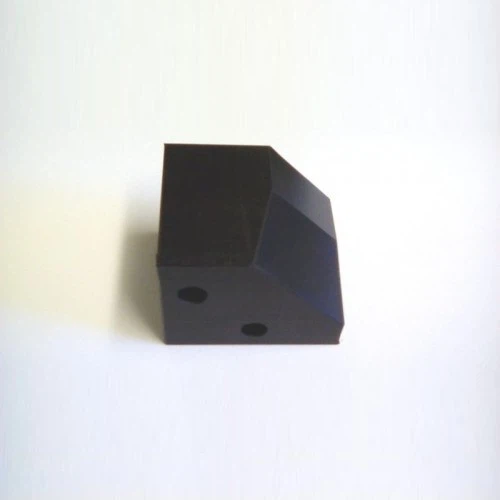

Secondly, tungsten has better mechanical properties than lead. It is harder and more durable, which means that tungsten - based components can withstand more wear and tear during normal use. This is particularly important in applications such as anti - scatter grids and collimators, where the components need to maintain their shape and performance over time.
In terms of radiation absorption, tungsten is comparable to lead for many X - ray energies. Although lead has a slightly higher atomic number (Z = 82), the difference in absorption efficiency is not significant enough to outweigh the advantages of tungsten in terms of safety and mechanical properties.
Future Developments
As medical imaging technology continues to evolve, the role of tungsten in scatter reduction is likely to become even more important. With the increasing demand for higher - quality images and lower radiation doses, there is a need for more advanced scatter reduction devices.
One area of future development is the optimization of the design of tungsten - based scatter reduction components. For example, researchers are exploring new grid designs that can further improve the scatter rejection ratio while maintaining a high transmission of primary X - rays. Computational modeling and simulation techniques are being used to predict the performance of different grid geometries and materials, allowing for more efficient design and development.
Another area of interest is the integration of tungsten into emerging imaging modalities. For example, in molecular imaging techniques such as positron emission tomography (PET) and single - photon emission computed tomography (SPECT), scatter radiation can also be a problem. Tungsten may have potential applications in these modalities, either in the form of shielding or as part of collimation systems.
In addition to its use in diagnostic imaging, tungsten also has applications in Tungsten for Nuclear Medicine. Nuclear medicine procedures involve the use of radioactive tracers, and scatter radiation can affect the accuracy of the imaging results. Tungsten - based collimators and shielding can help to improve the image quality and reduce the background noise in nuclear medicine imaging.
Conclusion
Tungsten plays a vital role in reducing scatter radiation in medical imaging. Its unique physical properties, including a high atomic number and density, make it an excellent material for scatter reduction devices such as anti - scatter grids, collimators, and shielding components. Compared to other materials such as lead, tungsten offers advantages in terms of safety and mechanical properties.
As a supplier of Tungsten for Medical Imaging, we are committed to providing high - quality tungsten products that meet the strict requirements of the medical industry. Our tungsten materials are carefully manufactured to ensure consistent performance and reliability. If you are interested in learning more about our products or have specific requirements for scatter reduction in your medical imaging applications, we invite you to contact us for procurement and further discussions. We look forward to working with you to improve the quality of medical imaging and reduce the radiation dose to patients and medical staff.
References
- Bushberg, J. T., Seibert, J. A., Leidholdt, E. M., & Boone, J. M. (2012). The essential physics of medical imaging. Lippincott Williams & Wilkins.
- Johns, H. E., & Cunningham, J. R. (1983). The physics of radiology. Charles C Thomas.
- Bissonnette, J. P. (2007). Principles of radiologic imaging: an art and a science. Saunders Elsevier.
